The cardiac conduction system is the electrical pathway of the heart that includes in order the SA node AV node bundle of His bundle branches and Purkinje fibers. Easily learn the conduction system of the heart using this step-by-step labeled diagram.

4 Posterior View Of The Human Heart Download Scientific Diagram
Day 1 Day 2 Day 3 By Region.

. Quiz yourself with the labeled views. Body cavity labeled diagram of organs they contain membranes and lateral views. Crayfish Dissection Virtual Crayfish Dissection Cornell Virtual Crayfish Dissection Penn State By Day.
3 µg of GFP plasmid overexpressed in mouse cardiomyocytes whole cell lysate with BSA for 1 hour at room temperature Lane 2. A Anterior view of the external heart C 2019 Pearson Education. Please help BlueLink grow by filling out this 2 minute survey to help us understand our users.
Midway is the central compartment of the thoracic cavitySurrounded by loose connective tissue it is an undelineated region that contains a group of structures within the thorax namely the heart and its vessels the esophagus the trachea the phrenic and cardiac nerves the thoracic duct the thymus and the lymph nodes. External Anatomy Internal Anatomy By Topic. The major changes that are made by the body occur at the first breath in the case of heart and lung functions and up to weeks after birth such as the livers enzyme synthesis.
Skeletal Integumentary Cardiovascular Muscular Endocrine Nervous Reproductive Respiratory Excretory Digestive You must create a series of labeled drawings. Learn about pacemaker cells and cardiac ac. 1 µg of GFP plasmid.
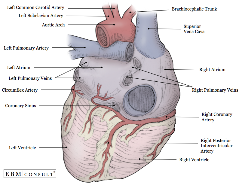
Ventral dorsal cranial spinal vertebral thoracic pleural pericardial mediastinum abdominopelvic abdominal and pelvic cavities explained. Anti-GFP antibody ab13970 at 12000 dilution Diluent 1x TBS 4 hours at 4C Lane 1. Aortc arch Ligamentum arteriosum Left pulmonary artery Left pulmonary ve ns Auricle of left atrium Circumflex artery Left coronary artery in atrioventricular sulcus Great cardiac vein Left ventricle Anterior interventricular artery in anterior interventricular sulcus Apex.
The mediastinum from Medieval Latin. Body cavity definitions and subdivisions in tables and charts. The foramen ovale becomes the fossa ovalis as the foramen closes while edge of the septum secundum in right atrium becomes anulus ovalis so the depression beneath it becomes the fossa ovalis.
Two granular areas referred to as Ig1 and Ig2 insular lobe granular areas found in the dorsal posterior insula and a dysgranular area labeled Id1 d for dysgranular area found in the. The only study to comprehensively analyze the cytoarchitecture of the human posterior insula using an observer-independent approach points to the existence of three distinct areas therein. Neurons in the developing central nervous system and brain congregate in layers or neighborhoods fitting into an alignment that will dictate their function.
2 µg of GFP plasmid overexpressed in mouse cardiomyocytes whole cell lysate with BSA for 1 hour at room temperature Lane 3.
Heart Posterior View Diagram Quizlet

Anterior And Posterior View Of Heart Human Anatomy Picture Cardiac Anatomy Human Heart Anatomy

Posterior View Of The Heart Diagram Quizlet

Posterior View Of The External Heart Diagram Quizlet
Anatomy Heart External

4 Posterior View Of The Human Heart Download Scientific Diagram

Posterior View Of The Heart Heart Anatomy Heart Diagram Anatomy

Heart Anatomy Labelled Illustration Stock Image C043 4821 Science Photo Library
0 comments
Post a Comment